
Written by: Dr. Jacquie Jacob, University of Kentucky
For anyone interested in raising chickens for eggs, whether for eating or incubation, an understanding of the female avian reproductive system is essential for recognizing problems that may occur and taking action to correct them.
The avian reproductive system is designed to accommodate the risks associated with being a bird. Other than birds of prey (such as hawks, eagles, and falcons), most birds are prey. Being close to the bottom of the food chain, birds require unique strategies for reproducing that also allow them to retain the ability to fly. For most birds, these unique strategies include producing many offspring and tending to the needs of the offspring for only a short period of time. The amount of time that birds devote to caring for their offspring depends on whether they are precocial or altricial birds, with the latter requiring more post-hatch parental care. Another reproductive strategy of birds is to produce offspring that develop outside the mother’s body in eggs. All the nutrients needed for an embryo to fully develop are provided in the egg before it is laid. It is for this reason that eggs are so nutritious for humans.
Poultry lay eggs in clutches. A clutch is a group of eggs laid by a hen on consecutive days. After laying a clutch, a hen has a rest period of about a day or more and then lays another clutch. Clutch sizes are species- and breed-specific. For commercial egg layers, clutch size is typically large. Clutch size, as well as the number of clutches laid in a hen’s laying cycle, varies by species, but the principle is the same across species.
An overview of the female chicken reproductive system helps explain why hens lay eggs in clutches. The reproductive system of a chicken hen is made up of two parts: the ovary and the oviduct. Ova (yolks) develop in the ovary. When an ovum (singular of ova) has matured, it is released from the ovary into the oviduct. This release of the ovum is ovulation. In the oviduct, glands secrete substances that form other parts of the egg, such as the albumen (egg white) and the shell. The total time a hen’s body takes to transform a yolk into a fully developed egg and lay that egg is about 25 to 26 hours. Typically, about 30 to 75 minutes after a hen lays an egg, the ovary releases the next ovum. However, the female chicken reproductive system is sensitive to light exposure, especially the number of hours of light in a day. In chicken hens, ovulation usually occurs under normal daylight conditions and almost never after 3:00 p.m. So, when a hen lays an egg too late in the day, the next ovulation occurs the following day, and the hen has a day when it does not lay an egg.
PARTS OF THE FEMALE CHICKEN REPRODUCTIVE SYSTEM
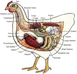
As stated, the female chicken reproductive system is made up of the ovary and the oviduct. (Figure 1 shows the female chicken reproductive system, and Figure 2 shows the location of the reproductive system in the body.) In almost all species of birds, including poultry, only the left ovary and oviduct are functional. Although the female embryo has two ovaries, only the left one develops. The right one typically regresses during development and is nonfunctional in the adult bird. (There have been cases in which the left ovary has been damaged and the right one has developed to replace it.)

OVARY
The ovary (shown in Figure 3) is a cluster of developing ova, and is located midway between the neck and the tail of the bird and attached at the back. The ovary is fully formed when a pullet chick hatches but is very small until the chick reaches sexual maturity. At hatch, a pullet chick has tens of thousands of ova, or potential eggs that theoretically could be laid, although most never develop to the point of ovulation. The maximum number of eggs a hen can lay is determined when it hatches because no new ova form after the chick hatches.

OVIDUCT
When ovulation occurs, the ovum (yolk) enters the oviduct. The oviduct is a twisted tube that is 25 to 27 inches long when fully developed and is divided into five major sections. These sections are the infundibulum, magnum, isthmus, shell gland, and vagina.
The first part of the oviduct, the infundibulum (or funnel) is 3 to 4 inches long and engulfs the ovum released from the ovary. The term funnel is an inaccurate name for this section because it suggests that the infundibulum is waiting for the yolk to fall into it, which is not the case. Instead, the released yolk stays in place, and the muscular infundibulum moves to surround it. The yolk remains in the infundibulum for 15 to 17 minutes. Fertilization, if it is going to occur, takes place in the infundibulum.
The next section of the oviduct is the magnum. At 13 inches long, it is the largest section of the oviduct, as its name implies (magnum being the Latin word for “large”). The yolk remains here 3 hours, during which time the thick albumen (egg white) forms.
The third section of the oviduct is the isthmus, which is 4 inches long. The isthmus, as its name implies, is slightly constricted (the term isthmus referring to a narrow strip of land joining two larger tracts of land). The isthmus is where the inner and outer shell membranes form. The developing egg remains here for 75 minutes.
The next section of the oviduct is the shell gland (or uterus), which is 4 to 5 inches long. In this section, the shell forms on the egg. The shell largely is made of calcium carbonate. The hen’s body mobilizes 8 to 10 percent of body calcium from its bones to make the egg’s shell. Bone calcium provides 47 percent of the calcium required to make a shell, and the hen’s diet provides the remainder. Pigment deposition, if there is any, occurs in the shell gland. The egg remains here for 20 or more hours.
The last part of the oviduct is the vagina, which is about 4 to 5 inches long. The vagina does not really play a part in egg formation but is important in the laying of the egg. The vagina is made of muscle that helps push the egg out of the hen’s body. The bloom, or cuticle, forms on the egg in the vagina prior to oviposition (the laying of the fully formed egg). The egg travels through the oviduct small end first but turns in the vagina and comes out large end first.
Near the junction of the shell gland and the vagina are deep glands known as sperm host glands that can store sperm for long periods of time, typically 10 days to 2 weeks. (One of the unique things about birds is that the sperm remain viable at body temperature.) When a hen lays an egg, sperm can be squeezed out of these glands into the oviduct and then can migrate to the infundibulum to fertilize an ovum.
EGG IRREGULARITIES
Various events can occur during reproduction that causes irregularities in eggs. Some of these irregularities affect the quality of the egg or consumer acceptance of the egg.
If the vitelline membrane surrounding the yolk becomes damaged, pale spots or blotches develop on the yolk. This irregularity is referred to as mottling. Although the appearance of the yolk has changed, there is no effect on the egg’s nutritional value and typically the mottling is not noticed by consumers. A high incidence of yolk mottling, however, adversely affects consumer acceptance. The use of cottonseed meal (which contains gossypol) and sorghum (which contains tannin) in the diet can increase the incidence of mottling. A calcium-deficient diet also has this effect.
Occasionally, a hen produces double-yolked eggs. This phenomenon can be related to hen-age, but genetic factors also are involved. Young hens sometimes release two yolks from the ovary in quick succession. Double-yolked eggs are typically larger in size than single-yolked eggs. Double-yolked eggs are not suitable for hatching as they typically have inadequate nutrients and space available for two chicks to fully develop and hatch. It has happened, but it is rare..
It is rare, but not impossible, for a young hen to produce an egg with no yolk at all. Yolk-less eggs (sometimes referred to as pullet eggs) are usually formed when a bit of tissue is sloughed off the ovary or oviduct. The tissue stimulates the secreting glands of the different parts of the oviduct and a yolk-less egg results.
Even rarer is an egg within an egg. This occurs when an egg nearly ready to be laid reverses direction, moves up the oviduct and encounters another egg in the process of forming. A new layer of albumen, new membranes, and a new shell form around the first egg, resulting in an egg inside an egg. Such eggs are so rare that no one knows exactly why they happen.
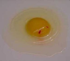
Other egg problems common when people raise their own chickens are blood spots (as shown in Figure 4) and meat spots. Blood spots are normally found on or around the yolk. The main cause of a blood spot is a small break in one of the tiny blood vessels around the yolk that occurs when the yolk is ovulated. High levels of hen activity during the time of ovulation can increase the incidence of blood spots. Meat spots are usually brown in color and are more often associated with the egg white. They form when small pieces of the wall of the oviduct are sloughed off while the developing egg is passing through. In commercial operations, eggs with blood spots and meat spots typically are identified during candling and removed (see Figure 5). It is rare, therefore, to find eggs with these irregularities in grocery stores. The incidence of blood spots is higher in brown-shelled eggs, and identifying blood spots when candling eggs with darker-colored shells is difficult.


Occasionally, a hen lays an egg without a shell. A shell-less egg feels like a water balloon. The shell membranes form around the yolk and egg white, but the egg somehow bypasses the shell-forming mechanism, and the shell is not completely deposited. The occurrence of the occasional shell-less egg is not necessarily an indication of health problems. If the incidence increases, however, a nutrition problem, primarily a deficiency of calcium, phosphorus, and/or vitamin D, may exist. If the condition persists, a veterinarian should examine the hen. Infectious bronchitis and egg drop syndrome also have been known to cause an increase in shell-less eggs.
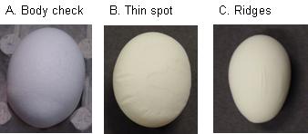
Other problems can occur when an egg’s shell is developing. The most obvious relates to shell texture. Occasionally, the shell becomes damaged while the egg is in the shell gland and is repaired before the hen lays the egg. This repair results in what is known as a body check (see Figure 6). Occasionally, thin spots in the shell or ridges form (see Figure 6). These shells are weaker than those of normal eggs, so eggs with thin spots are removed during inspection of table eggs and should not be used as hatching eggs.
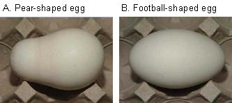
The second category of problems relates to abnormal shape (see Figure 7). Eggs with abnormal shapes do not fit well into a typical egg carton or are more likely to break during transport, so they are removed during egg inspection and are not normally sold in stores. Hatching eggs also should have the typical egg shape. With many abnormally shaped eggs, it is not clear which is the large end, and eggs should be incubated large end up. Also, such eggs may not properly fit in the egg trays.